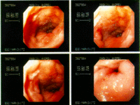
Endoscopy means looking inside and typically refers to looking inside the body for medical reasons using an instrument called an endoscope. Endoscopy can also refer to using a borescope in technical situations where direct line-of-sight observation is not feasible.
Contents[hide] |
Overview
Endoscopy is a minimally invasive diagnostic medical procedure that is used to assess the interior surfaces of an organ by inserting a tube into the body. The instrument may have a rigid or flexible tube and not only provide an image for visual inspection and photography, but also enable taking biopsies and retrieval of foreign objects. Endoscopy is the vehicle for minimally invasive surgery, and patients may receive conscious sedation so they do not have to be consciously aware of the discomfort.
Many endoscopic procedures are considered to be relatively painless and, at worst, associated with mild discomfort; for example, in esophagogastroduodenoscopy, most patients tolerate the procedure with only topical anaesthesia of the oropharynx using lignocaine spray. [1] Complications are not common (only 5% of all operations)[citation needed] but can include perforation of the organ under inspection with the endoscope or biopsy instrument. If that occurs open surgery may be required to repair the injury.
Components
An endoscope can consist of
- a rigid or flexible tube
- a light delivery system to illuminate the organ or object under inspection. The light source is normally outside the body and the light is typically directed via an optical fiber system
- a lens system transmitting the image to the viewer from the fiberscope
- an additional channel to allow entry of medical instruments or manipulators
Uses
Endoscopy can involve
- The gastrointestinal tract (GI tract):
- esophagus, stomach and duodenum (esophagogastroduodenoscopy)
- small intestine
- colon (colonoscopy,proctosigmoidoscopy)
- Bile duct
- endoscopic retrograde cholangiopancreatography (ERCP), duodenoscope-assisted cholangiopancreatoscopy, intraoperative cholangioscopy
- The respiratory tract
- The nose (rhinoscopy)
- The lower respiratory tract (bronchoscopy)
- The urinary tract (cystoscopy)
- The female reproductive system
- The cervix (colposcopy)
- The uterus (hysteroscopy)
- The Fallopian tubes (Falloscopy)
- Normally closed body cavities (through a small incision):
- The abdominal or pelvic cavity (laparoscopy)
- The interior of a joint (arthroscopy)
- Organs of the chest (thoracoscopy and mediastinoscopy)
- During pregnancy
- The amnion (amnioscopy)
- The fetus (fetoscopy)
- Plastic Surgery
- Panendoscopy (or triple endoscopy)
- Combines laryngoscopy, esophagoscopy, and bronchoscopy
- Non-medical uses for endoscopy
- The planning and architectural community have found the endoscope useful for pre-visualization of scale models of proposed buildings and cities (architectural endoscopy)
- Internal inspection of complex technical systems (borescope)
- Endoscopes are also a tool helpful in the examination of improvised explosive devices by bomb disposal personnel.
- The FBI uses endoscopes for conducting surveillance via tight spaces.
History
The first endoscope, of a kind, was developed in 1806 by Philip Bozzini with his introduction of a "Lichtleiter" (light conductor) "for the examinations of the canals and cavities of the human body". However, the Vienna Medical Society disapproved of such curiosity. An endoscope was first introduced into a human in 1822 by William Beaumont, an army surgeon at Mackinac Island, Michigan[citation needed]. The use of electric light was a major step in the improvement of endoscopy. The first such lights were external. Later, smaller bulbs became available making internal light possible, for instance in a hysteroscope by Charles David in 1908[citation needed]. Hans Christian Jacobaeus has been given credit for early endoscopic explorations of the abdomen and the thorax with laparoscopy (1912) and thoracoscopy (1910)[citation needed]. Laparoscopy was used in the diagnosis of liver and gallbladder disease by Heinz Kalk in the 1930s[citation needed]. Hope reported in 1937 on the use of laparoscopy to diagnose ectopic pregnancy[citation needed]. In 1944, Raoul Palmer placed his patients in the Trendelenburg position after gaseous distention of the abdomen and thus was able to reliably perform gynecologic laparoscopy[citation needed]. In the early 1950s Harold Hopkins designed a “fibroscope” (a flexible bundle of glass fibres), which proved useful both medically and industrially, paving the way for modern fibre optic cables. Hopkins refined his ideas with a German manufacturer, resulting in a design breakthrough in 1967 which enabled surgeons to view and photograph inaccessible areas of the body in detail.
Risks
- Infection
- Punctured organs
- Allergic reactions due to Contrast agents or dyes (such as those used in a CT scan)
- Over-sedation
After the endoscopy
After the procedure the patient will be observed and monitored by a qualified individual in the endoscopy or a recovery area until a significant portion of the medication has worn off. Occasionally a patient is left with a mild sore throat, which promptly responds to saline gargles, or a feeling of distention from the insufflated air that was used during the procedure. Both problems are mild and fleeting. When fully recovered, the patient will be instructed when to resume his/her usual diet (probably within a few hours) and will be allowed to be taken home. Because of the use of sedation, most facilities mandate that the patient is taken home by another person and not to drive on his/her own or handle machinery for the remainder of the day.
Recent developments
With the application of robotic systems, telesurgery was introduced as the surgeon could operate from a site physically removed from the patient. The first transatlantic surgery has been called the Lindbergh Operation.


1 comment:
smart outsourcing solutions is the best outsourcing training
in Dhaka, if you start outsourcing please
visit us: It training institute in dhaka
Post a Comment